The spine, or vertebral column, is a crucial structure that supports the body, enables movement, and protects the spinal cord. It's composed of 33 individual bones called vertebrae, stacked upon each other.
When viewed from the side, a healthy adult spine exhibits a natural S-shaped curve. The neck (cervical) and lower back (lumbar) regions curve slightly inward (concave), while the mid-back (thoracic) and tailbone (sacral) regions curve gently outward (convex). These curves act as shock absorbers, helping to maintain balance and allow for a wide range of motion.
The spine is stabilized and moved by two main muscle groups: extensors and flexors. Extensors, located on the back, enable us to stand and lift objects. Flexors, including the abdominal muscles in the front, allow us to bend forward and control the arch of the lower back.
7 vertebrae (C1-C7) that support the head and allow for a wide range of motion. The first two vertebrae, the atlas (C1) and axis (C2), are specialized for head movement.
12 vertebrae (T1-T12) that hold the rib cage and protect the heart and lungs.
5 vertebrae (L1-L5) that bear the weight of the body. These are the largest vertebrae.
5 fused vertebrae that connect the spine to the hip bones.
4 fused bones that form the tailbone.
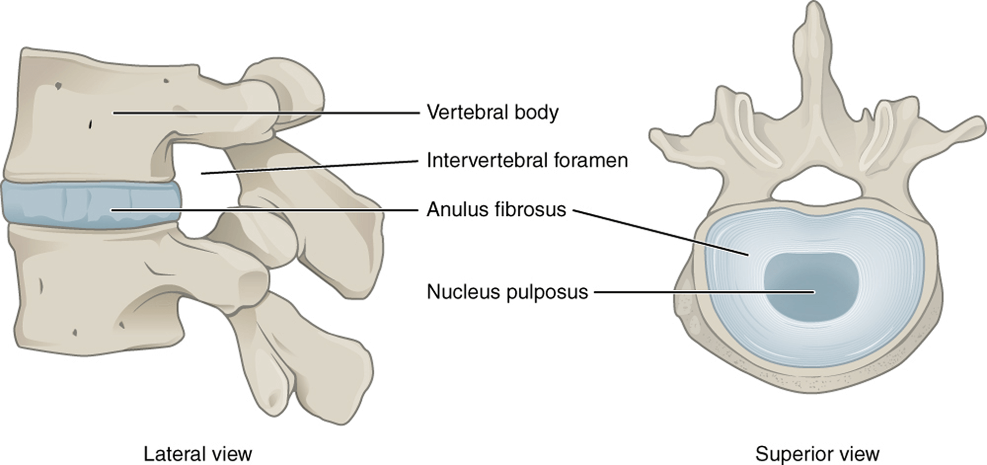
Between each vertebra lies an intervertebral disc, which acts as a cushion and prevents the bones from rubbing together. The disc has a tough outer ring (annulus) and a gel-filled center (nucleus).
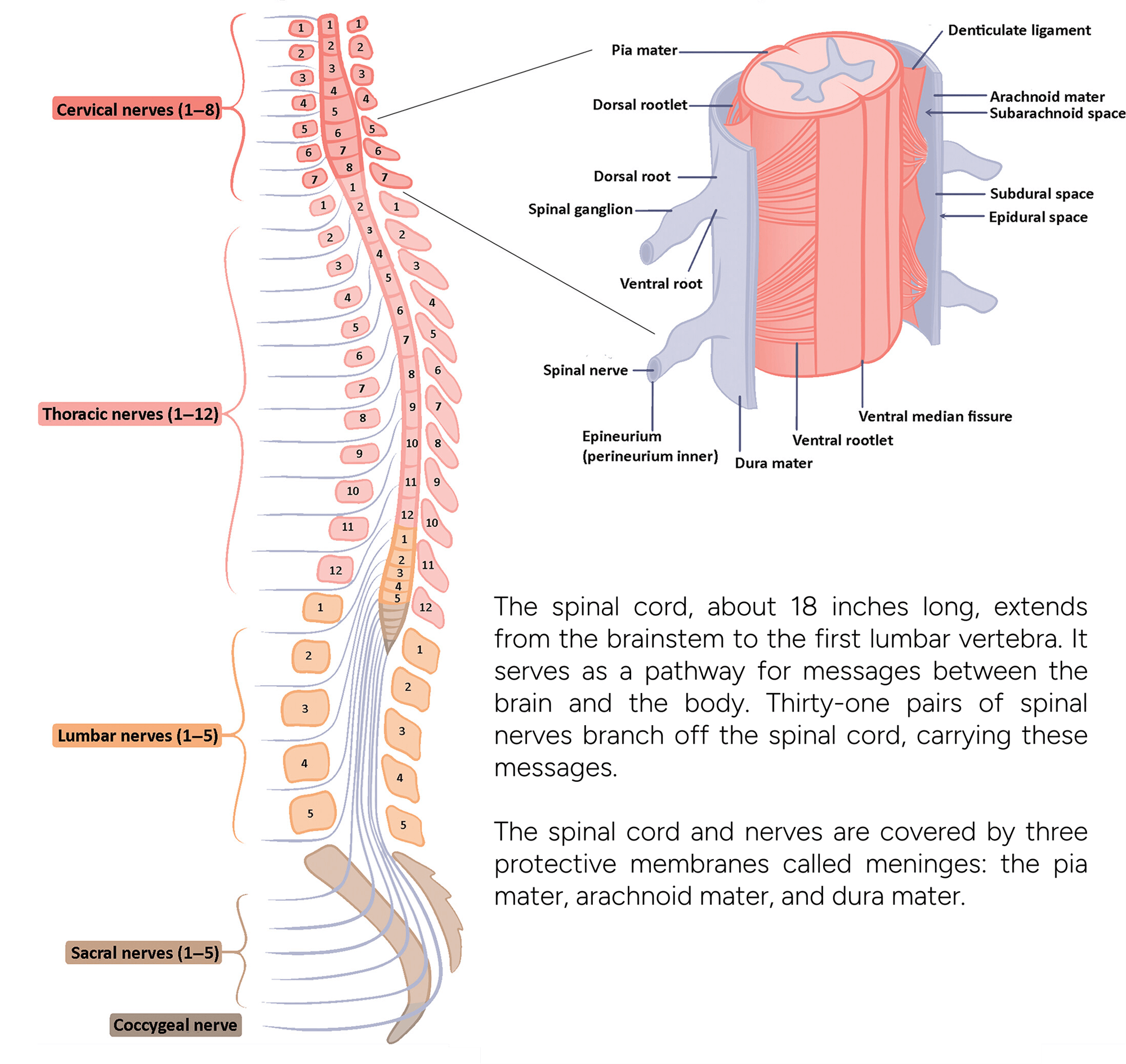
The back of each vertebra features bony projections that form the vertebral arch. The spinal cord, along with fat, ligaments, and blood vessels, runs through the spinal canal within this arch. Spinal nerves exit the spinal cord through openings called intervertebral foramina.
Facet joints allow for back motion. Each vertebra has four facet joints that connect it to the vertebrae above and below.
Ligaments, strong fibrous bands, hold the vertebrae together, providing stability and protecting the discs.